by Nirvan Garg, 11
Mosses provide a habitat to many microscopic species, including tardigrades (water bears) – famous for being incredibly resilient, such that they can survive space. Other such residents include Nematodes, Daphnia, Algae, etc.
With the current weather being remarkably rainy, moss has become quite abundant, and this is the perfect opportunity to observe some of these creatures under a microscope.
Apparatus:
• Spatula
• Petri dish
• Optical microscope
• Glass slide
• Coverslip
• Dropper
• Gloves
• Purified water (NOT DEIONISED OR DISTILLED)
• Hand sanitiser (for sterilisation of work environment afterwards)
• A coin or small opaque circular object (to place on the illuminator, to enable dark field illumination)
Procedure:
• Locate a growth of moss. Use the spatula to scrape some moss into the Petri dish. Ensure there is a substantial amount of sample.
• Use the dropper to add a small volume of purified water to the Petri dish. It is important not to use deionised or distilled water, as we do not want to give any of the microanimals osmotic shock. It is also important that the amount of water added is not enough to submerge the moss sample.
• Swirl the contents of the Petri dish lightly or mix by pumping the liquid in the dropper. Leave to rest and hydrate for 24hrs.
• After 24hrs, take some liquid from the Petri dish in the dropper and place one drop on a glass slide. Carefully add a coverslip and view the slide under the microscope.
• GOING FURTHER: Place the coin (or small opaque circular object) in the centre of the illuminator of the microscope. This blocks the light coming from the centre of the illuminator, and as a result only highly scattered light enters the objective lens, as most light does not pass straight through the sample into the lens. This means that the background is dark. The advantage of this is that because tardigrades (etc.) are almost translucent, they can be hard to see under brightfield illumination. With a dark background and dark field illumination, the outlines of the creatures show up bright white. However, this is an additional step and most creatures are usually visible using bright field illumination as well.
This procedure can be utilised for most liquid samples containing living specimens that need to be examined. For
example, the same process can be repeated with pond water.
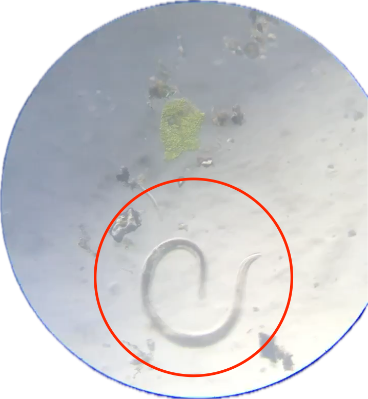
moss sample. Image quality is poor as it is
an extract from a video, where the nematode
can clearly be seen swimming in the FOV.
IMAGE CREDIT: AUTHOR
Leave a Reply